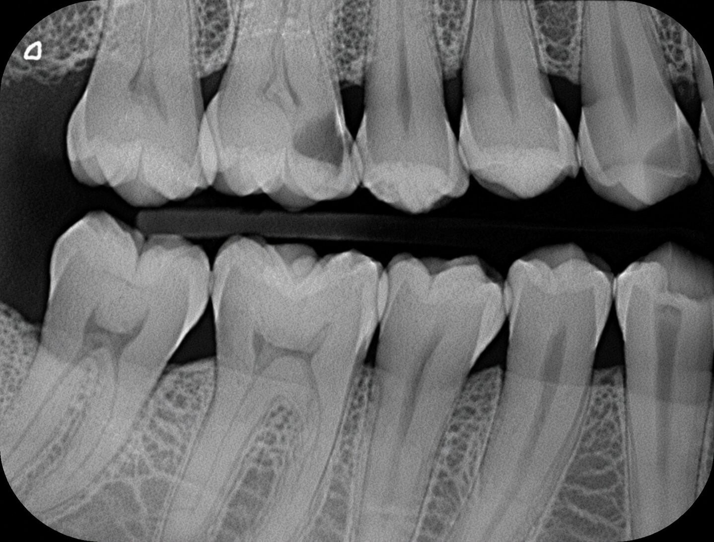
You already know the normal landmarks (Lesson 02) and the radiopaque/radiolucent reflex (Lesson 10). Now the exam adds the twist: real conditions — caries, apical pathology, periodontal disease, implants, missing teeth, extra teeth, the jaw joint — that have to be told apart from normal anatomy, on sight.
7 conditions~8 min★ Domain I — Purpose & Technique
▶ Watch · 2:08
Watch first — landmark or lesion, in a little over two minutes.
1The problem
The exam loves the look-alike.
A small round radiolucency at a premolar apex could be the mental foramen — completely normal — or it could be a periapical abscess. Same size, same location, same dark shadow. The difference is everything: symptoms, borders, and whether it shows up on both sides.
This lesson is a tour of the conditions DANB actually names — caries, apical pathology, periodontal disease, implants, edentulous arches, dental anomalies, and the TMJ — with the one habit that sorts every one of them: is it symmetric and well-defined (normal), or off-center, irregular, and tied to symptoms (pathology)?
2The instrument
Pick a condition. See it.
Tap a condition. Watch the schematic mark where it shows up and how it reads — then read what makes it different from a normal landmark.
That single question — does it show up on both sides, and does anything hurt? — sorts most of the exam's "is this normal?" questions. A textbook example: a small round radiolucency at a premolar apex with no symptoms is almost always the mental foramen, not an abscess. Bilateral, symmetric findings default to normal anatomy; asymmetric findings tied to pain, swelling, or mobility default to pathology.
4The essentials
Everything on one card.
Caries
Radiolucent — decay removes mineral. Interproximal caries is the classic bitewing finding; a triangular shadow pointing toward the pulp is the giveaway shape.
Apical pathology
A radiolucent area at a root apex — abscess, granuloma, or cyst. All three can look similar on a single image; a cyst tends to have a thin radiopaque (corticated) border, an abscess is often more diffuse. Symptoms and history tell them apart, not the image alone.
Periodontal disease
Reads as loss of crestal bone height below where it should sit, plus a widened periodontal ligament space or lost lamina dura. A vertical bitewing is the best single view for bone levels.
Dental implants
A dense, threaded radiopaque fixture embedded in bone — nothing else in the mouth looks like it. Best evaluated with a periapical, which shows the full length and surrounding bone.
Edentulous arches
No tooth to anchor a normal bitewing/periapical setup — the ridge itself (resorbed, smoother) becomes the landmark. Panoramic is typical for a full edentulous survey.
Dental anomalies
Supernumerary (extra) teeth are best screened on a panoramic, then localized with a periapical/occlusal. Dilaceration (a sharply bent root) is a true anomaly too — not an angulation error.
TMJ
The jaw joint — condyle seated in the glenoid fossa. A panoramic gives a limited view; a dedicated TMJ series or CBCT is needed to properly evaluate joint position and pathology.
5Beat the exam
How DANB will actually ask this.
It describes a finding and wants you to say "normal" or "pathology" — or names a condition and wants the right image. Match the words:
"small round radiolucency at a premolar apex, no symptoms"→mental foramen — a normal landmark
"a finding on both sides, same spot"→normal anatomy — bilateral defaults to landmark
"evaluate crestal bone loss"→vertical bitewing
"screen for extra (supernumerary) teeth"→panoramic
"evaluate an implant / its surrounding bone"→periapical
6Check yourself
A periapical shows a small, round, symmetric radiolucency near the apex of a mandibular premolar. The patient has no pain or swelling. This is MOST likely:
The mental foramen. A round, well-defined periapical radiolucency near the mandibular premolars with no symptoms is the classic "landmark mistaken for pathology." True apical pathology usually comes with a symptom history and less regular borders.
6Check yourself
Which radiograph best evaluates crestal bone levels for periodontal disease?
Vertical bitewing. Turning the bitewing vertically captures more crown-to-bone height in one image, making it the best single view for reading periodontal bone loss.
6Check yourself
Supernumerary (extra) teeth are best initially detected using which image?
Panoramic. It surveys the whole dentition and both jaws in one image, making it the best screening tool for extra teeth. A periapical or occlusal can then localize a specific one.
Recap
The whole thing in 5 lines.
✓Ask one question first: symmetric & symptom-free (normal landmark) or off-center & symptomatic (pathology)?
✓Caries and apical pathology (abscess/granuloma/cyst) both read radiolucent — location and shape separate them from landmarks like the mental foramen.
✓Periodontal disease = crestal bone loss, best read on a vertical bitewing.
✓Implants read as a dense threaded fixture (periapical to evaluate); edentulous arches lose the tooth landmark entirely (panoramic for a full survey).
✓Supernumerary (extra teeth) screen on panoramic; the TMJ needs more than a panoramic to fully evaluate.
Every finding asks the same question: landmark, or lesion? Symmetry and symptoms give you the answer.
You've now got the whole Domain I interpretation toolkit — normal anatomy, density, and the conditions that break the pattern. Next: whether the image itself is good enough to read at all.