Landmarks are the #1 thing test-takers say tripped them up — and they're heavily tested. But here's the secret: you don't memorize them one by one. You learn one rule, and the rest fall into place.
1 rule to learn20 landmarks~12 min★ The most-missed, most-tested topic on the RHS exam
▶ Watch · 2:21
Watch first — radiopaque materials, real images and real examples, in under two and a half minutes.
1Why it matters
You can't spot a problem if you don't know "normal."
Your whole job on an x-ray is to recognize when something looks wrong — a cavity, an abscess, an extra tooth. But you can only do that if you first know what the normal anatomy looks like. A dark spot might be a serious problem… or it might just be the mental foramen, which is supposed to be there. Knowing the landmarks is what lets you tell the difference.
And here's the part that makes it easy: whether a structure shows up white or black on the film isn't random. It follows one simple rule about what blocks x-rays and what lets them through. Learn the rule, and most of the "memorizing" disappears.
The whole thing in one sentence
Solid bone blocks x-rays and shows up white; holes and spaces let them through and show up black. The landmark's name usually tells you which.
In real terms: radiopaque = white (bone, metal); radiolucent = black (foramen, canal, sinus, fossa).
2Start with shadows
First, forget the anatomy — think about shadows.
Remember an x-ray isn't a photo — it's a shadow picture. The x-ray beam shoots through the head toward the sensor. Wherever something solid blocks the beam, fewer x-rays get through, and that spot on the image stays light/white. Wherever there's a gap — a hole, an air space, a hollow — the beam sails right through and darkens the sensor, so that spot turns black.
🔦
It's just "what blocks the light?"
Hold your hand in front of a flashlight pointed at a wall. Your solid hand makes a dark shadow — it blocked the light. The gaps between your fingers let light through — bright. X-rays work the opposite in color (blocked = white, not dark), but the idea is identical: solid stuff stops the beam, empty space lets it through. That single fact decides every landmark's color.
HAND (SOLID) BLOCKS LIGHT = DARK SHADOW
GAPS BETWEEN FINGERS LET LIGHT THROUGH = BRIGHT
The flashlight analogy, made literal: the solid hand blocks the light and casts a dark shadow — same as bone blocking x-rays. The gaps between the fingers let light straight through — same as a foramen, canal, or sinus letting x-rays through.
So before any anatomy words: solid = blocks the beam = shows white. Hollow = lets the beam through = shows black. Hold onto that — it's the whole lesson.
3The one rule
Bone → WHITE Hole / space → BLACK
Bone blocks x-rays → shows up white (radiopaque). Openings, canals, sinuses, fossae let x-rays through → show up black (radiolucent). The name usually tells you which.
4The two words
The naming trick.
The dental words for "white" and "black" sound fancy, but they just mean blocks-the-beam or lets-it-through:
Radiopaque
= white / light
The structure blocked the x-rays (it's dense — bone or metal), so the sensor stayed light.
Name tells you: process, ridge, nasal spine, tuberosity, septum, lamina dura — and the walls of any space. Plus all metal (fillings, crowns).
Radiolucent
= black / dark
The x-rays passed through (it's a gap or opening), so the sensor went dark.
Name tells you: foramen, canal, fossa, sinus, suture, space — anything that's an opening or hollow.
Here's the trick that does the heavy lifting: you're not learning Latin — you're learning what each word means. Learn the plain-English meaning, and the meaning tells you the color. Here are the exact words you'll see on the exam:
If the name means…It shows up…
🕳️ForamenA hole or opening in bone where nerves/vessels pass through. / canalA tube-like passage through bone for nerves and vessels. — "a hole"
→
Radiolucent (black)the beam goes through the opening
🥄FossaA shallow scooped-out depression in bone. / sinusA hollow air-filled cavity in bone. — "a hollow"
→
Radiolucent (black)less bone = beam passes through
⛰️ProcessA marked projection or bump of bone. / ridge / nasal spine / tuberosity — "a bump"
→
Radiopaque (white)extra solid bone blocks the beam
🧱SeptumA bony wall that divides two spaces. / suture / wall / lamina dura — "a wall"
→
Mixed — read carefullya bony wall = white; a suture (gap between bones) = black
One honest catch: suture is the exception that breaks the "wall" pattern — a suture is the thin gap between two skull bones, so it reads black (radiolucent). The median palatal suture is the classic example. Hover any dotted wordLike this! Tap or hover any dotted word for a plain-English definition. to refresh it.
5Sort it yourself
Sort each landmark: white or black?
Drag each landmark into the right bucket — Radiopaque (white) or Radiolucent (black). Use the naming trick: hole/space = black, bump/wall = white. Wrong answers bounce back so you can try again. (On a touchscreen, press and drag — or tap a chip, then tap a bucket.)
Drag each landmark into its bucket ↓
⬜
Radiopaque
white — blocked the beam (bone / metal)
⬛
Radiolucent
black — beam passed through (hole / space)
The landmarks
Remember: a hole, canal, sinus, or space lets the beam through (black). Bone and metal block it (white).
0 of 10 placed
Notice you didn't have to memorize a single one — you reasoned each from its name. That's the skill the exam is really testing.
6Every landmark
🔼 Maxillary — Anterior (front of upper jaw)
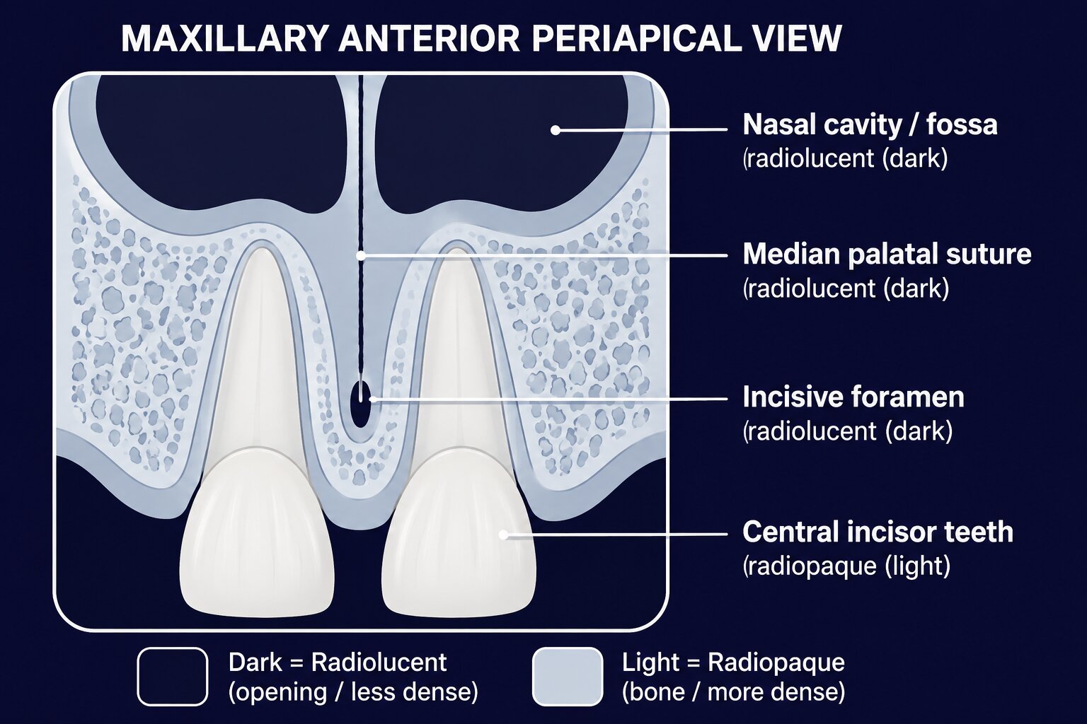
See it in place: the dark openings (nasal cavity, suture, incisive foramen) versus the light, dense teeth and bone. Match each label below to the picture.
Incisive foramen Radiolucent
Small oval black spot between the roots of the upper central incisors. (A foramen = a hole.)
Median palatal suture Radiolucent
Thin black vertical line between the upper centrals — the joint between the two palate halves.
Nasal cavity / fossa Radiolucent
Large black area above the upper incisors (the air space of the nose).
Nasal septum Radiopaque
White vertical bony wall dividing the nasal cavity into left and right.
Anterior nasal spine Radiopaque
White V-shaped point where the nasal floor meets the septum.
6Every landmark
🔼 Maxillary — Posterior (back of upper jaw)
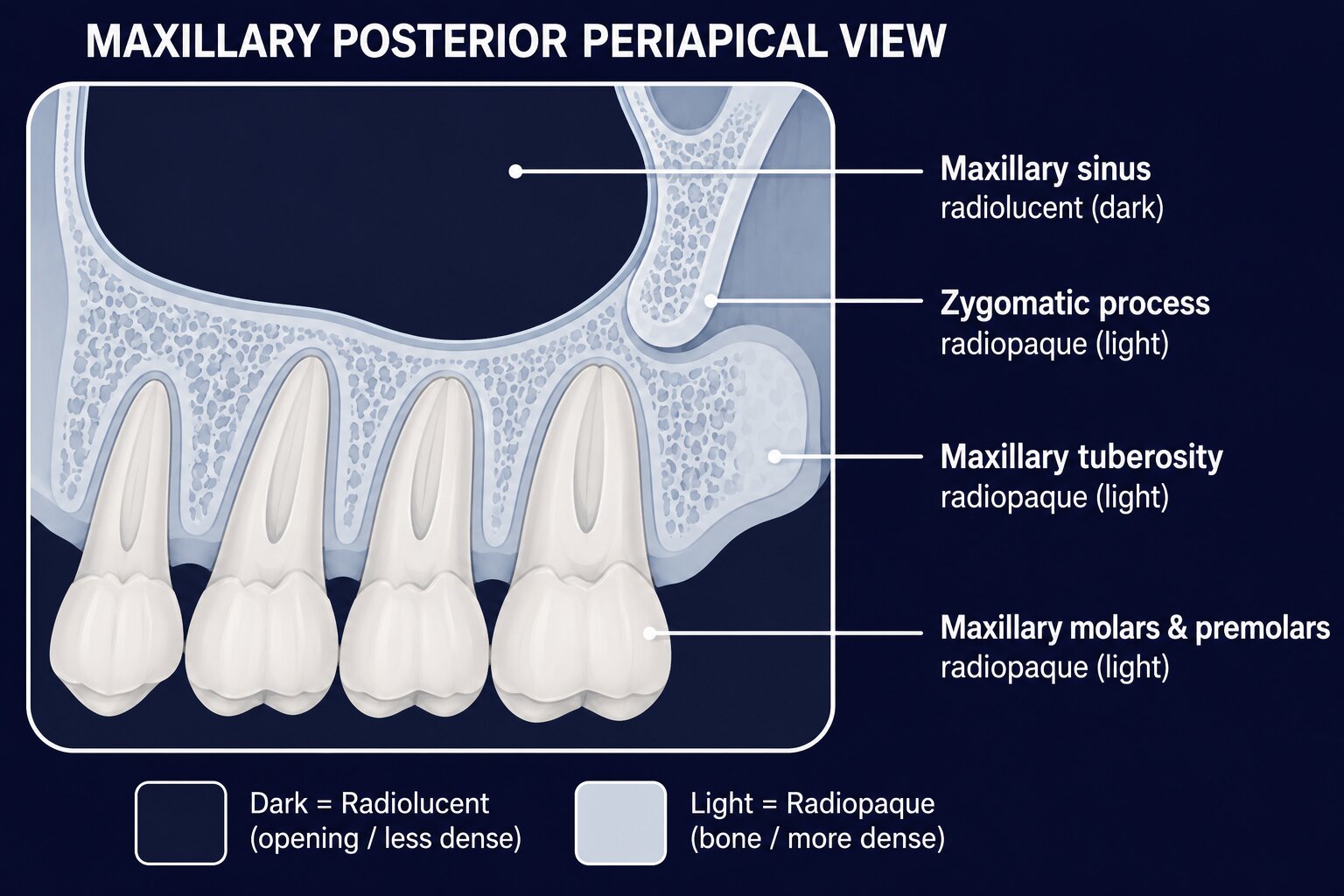
The big dark space above the roots is the maxillary sinus — the most-tested landmark here. Notice the dense, light bone of the zygomatic process and tuberosity around it.
Maxillary sinus Radiolucent
Large black air cavity above the upper premolar/molar roots, with a thin white bony border.
Zygomatic process Radiopaque
White J- or U-shaped band above the upper first molar (cheekbone attachment).
Maxillary tuberosity Radiopaque
White rounded bulge of bone behind the last upper molar.
Coronoid process Radiopaque
White triangle (it's part of the mandible) that drifts into the upper molar view when the mouth opens.
Inverted Y Radiopaque
White upside-down Y over the canine — where the sinus and nasal cavity walls cross.
6Every landmark
🔽 Mandibular — Anterior (front of lower jaw)
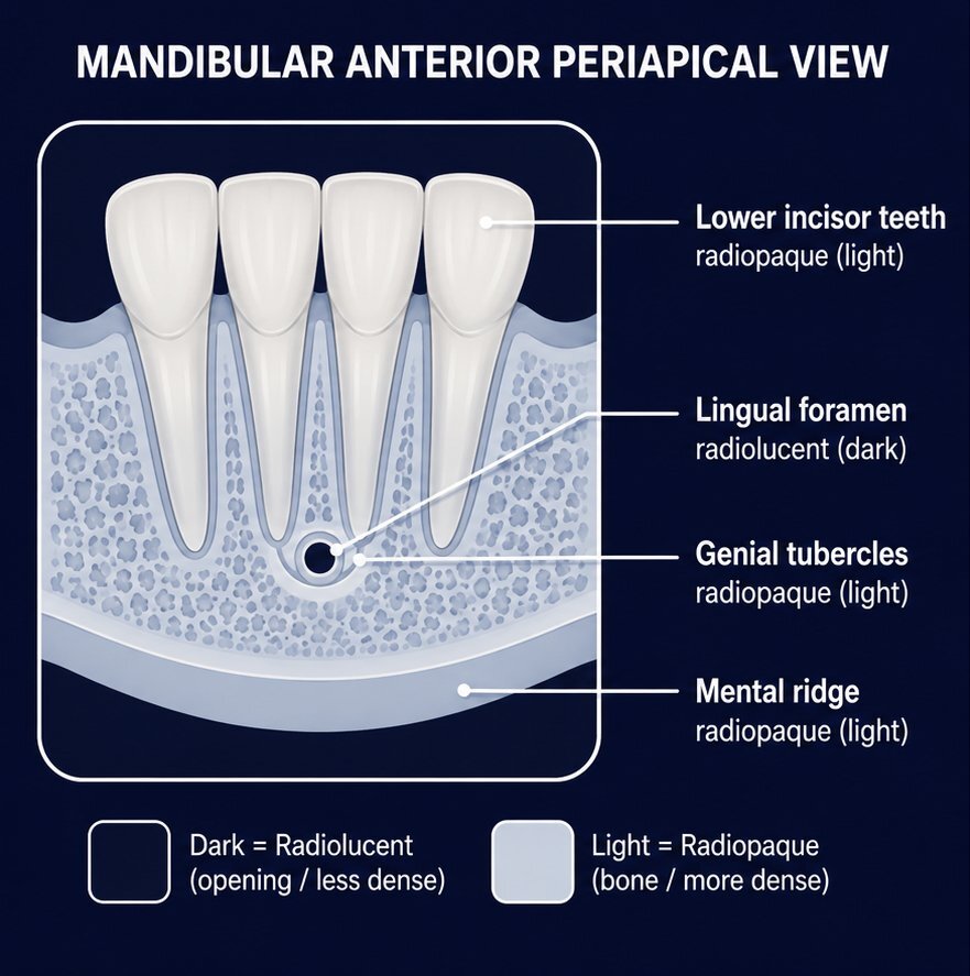
The lingual foramen is the tiny dark dot at the midline — ringed by the light genial tubercles. That ring-around-a-dot pattern is a classic exam clue.
Genial tubercles Radiopaque
White ring or mass of bone below the lower incisor roots (muscle attachment bumps).
Lingual foramen Radiolucent
Tiny black dot in the middle of the genial tubercles (a hole).
Mental ridge Radiopaque
White band sweeping up from premolar toward incisor region (a ridge).
Nutrient canals Radiolucent
Thin black vertical lines running up between the lower front teeth — tiny channels that carry blood vessels. Normal, and most visible in the anterior mandible. (A canal = a passage → black.)
6Every landmark
🔽 Mandibular — Posterior (back of lower jaw)
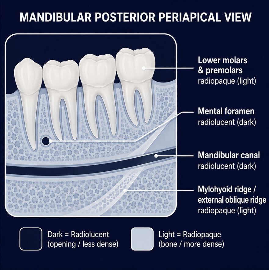
The mandibular canal is the dark band running below the roots between two thin white walls. The mental foramen is a separate dark dot near the premolars — easy to mistake for an abscess.
Mental foramen Radiolucent
Small round black spot near the lower premolar apices — often mistaken for an abscess!
Mandibular canal Radiolucent
Black band below the molar roots, outlined by two thin white walls (houses the nerve).
Mylohyoid ridge Radiopaque
White band running down-and-forward from the molar region (a ridge).
External/internal oblique ridge Radiopaque
White bands in the molar region (ridges of the ramus).
Submandibular fossa Radiolucent
Black scooped-out area below the mylohyoid ridge (holds a salivary gland).
Inferior border of the mandible Radiopaque
Thick, bright white band of dense cortical bone forming the bottom edge of the lower jaw — the densest normal landmark on a lower film. (Solid bone → white.)
6Every landmark
Don't panic at the list length. You're not memorizing 20 things — you're applying one rule 20 times. Foramen, canal, sinus, fossa, space, suture → black. Process, ridge, nasal spine, tuberosity, septum, wall, metal → white.
★
Two the exam hides in plain sight
The frontal sinus is a trap — you will never see it on an intraoral film. Of the paranasal sinuses, only the maxillary sinus lands on periapicals and bitewings. The frontal, ethmoid, and sphenoid sinuses sit too high and show up only on panoramic or other extraoral images. So if a question points at a sinus on an inside-the-mouth film, the answer is the maxillary sinus.
The maxillary sinus is also your mounting compass. It exists only in the upper jaw, so the moment you spot that big dark air space sitting above the roots, you know you are looking at a maxillary (upper) film — a fast orientation check when you mount.
7Beat the exam
How DANB will actually ask this.
Two flavors: "is this structure radiopaque or radiolucent?" and "what is this landmark?" (sometimes with a picture, sometimes described in words — and the wording can be wordier than your study guide). The rule handles both. The #1 trap to know cold:
"round dark spot near lower premolar roots"→Mental foramen (normal) — NOT an abscess or cavity
"dark area — is it pathology?"→First ask: is there a normal landmark there?
8Check yourself
On a periapical image, the maxillary sinus appears as which of the following?
A radiolucent (dark) area above the premolar/molar roots. A sinus is a hollow air space — the beam passes through, so it's black. The rule predicts it without memorizing.
8Classic trap
A small, round, dark (radiolucent) area is seen near the apex of a mandibular premolar on a routine radiograph. The patient has no symptoms. This most likely represents:
The mental foramen. A foramen is a normal hole in bone — it's radiolucent and sits right at the premolar apices. No symptoms + expected location = normal anatomy, not pathology. This is the most common landmark trap on the exam.
8Lock it in
Which of the following normal structures would appear RADIOPAQUE (white) on a dental image?
Lamina dura. It's the dense bony wall lining the tooth socket — solid bone blocks the beam, so it's white. The other three are a foramen (hole), a canal (passage), and a fossa (hollow) — all radiolucent.
8Bonus round
Which paranasal sinus is normally visible on an intraoral (inside-the-mouth) radiograph?
The maxillary sinus. It sits right above the upper premolar and molar roots, so it lands on periapicals and bitewings. The frontal, sphenoid, and ethmoid sinuses are too high and show only on panoramic or other extraoral films.
Recap
The whole thing in 4 lines.
✓An x-ray is a shadow: solid bone blocks the beam → white (radiopaque); holes & spaces let it through → black (radiolucent).
✓The name tells you the color: foramen / canal / sinus / fossa / space / suture = black; process / ridge / nasal spine / tuberosity / wall / metal = white.
✓Know the regions: maxillary & mandibular, anterior & posterior — but reason each landmark from the rule, don't rote-memorize.
✓The classic trap: a dark spot by the lower premolars is usually the mental foramen (normal), not pathology.
That's the whole lesson. On test day you won't memorize landmarks — you'll reason each one from a single rule.
Foramen, canal, sinus, fossa, space, suture read black; process, ridge, nasal spine, tuberosity, septum, wall and metal read white. Apply it once and a landmark you've never seen still gives itself away.