Every reading starts with one skill: is it radiopaque (light) or radiolucent (dark)? Get that reflex automatic and you stop falling for the handful of look-alikes the exam plants to fool you.
~7 min2 words to master★ Cervical burnout — the #1 caries impostor
▶ Watch · 1:57
Watch first — radiopaque vs. radiolucent in under two minutes.
1The one idea
Density is the whole game.
An x-ray image is a map of what stopped the beam. Dense things — enamel, bone, metal — absorb the x-rays, so little reaches the receptor and that spot stays radiopaque (light/white). Thin or empty things — soft tissue, air, a cavity where tooth used to be — let the beam through, so that spot goes radiolucent (dark).
That's it. Radiopaque = light = dense.Radiolucent = dark = less there. Caries reads dark because decay has removed mineral; metal reads bright white because nothing gets through it.
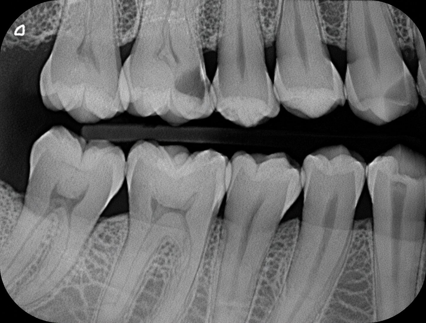
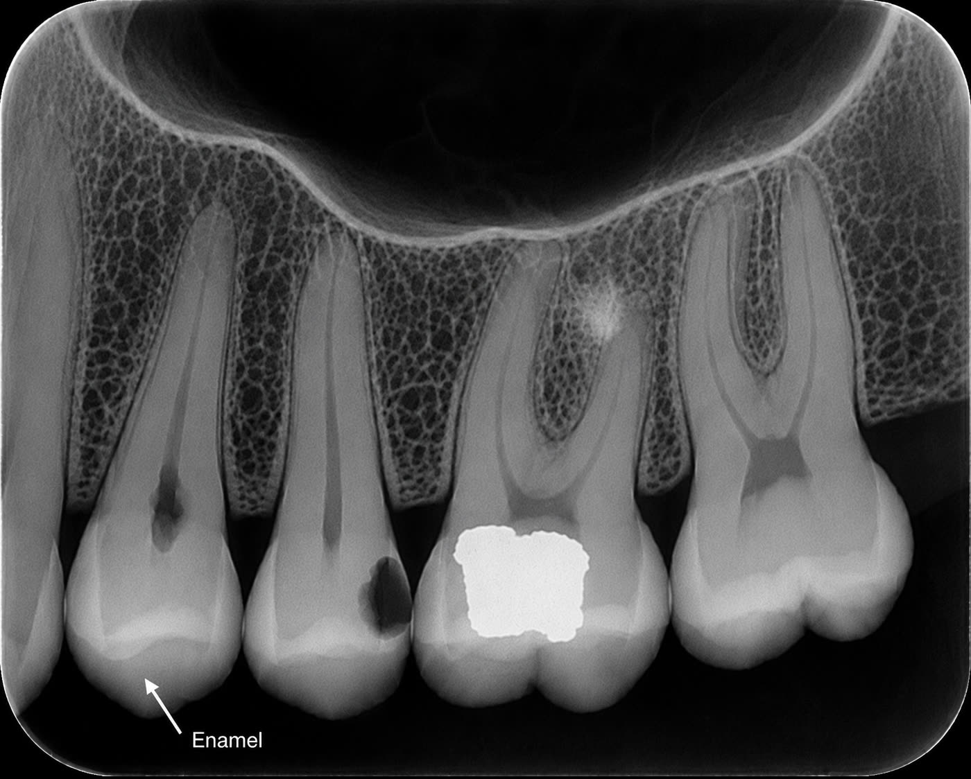
Interproximal caries
See it for real. On the proximal (side) surface of the tooth, that dark notch is interproximal caries — radiolucent because decay removed tooth structure. The bright white around it is intact enamel.
2The instrument
Light or dark?
Tap a structure or finding. Call it in your head first — radiopaque or radiolucent — then see the film go light or dark, and why.
The density sorter
Fig. 10.1 · interactive
The structure or finding
On the film
Radiopaque · light
Enamel
—
3The rule, in two lines
Opaque = light = dense Lucent = dark = gone
Say it until it's automatic. The densest things in the mouth — metal restorations — are the whitest; then enamel, then bone and dentin. The darkest are air and anything that has lost tooth structure — a cavity, the pulp, the thin PDL line.
4The whole scale
Every structure, sorted.
From the brightest to the darkest. If you can place a new finding on this scale, you can read it.
Structure / finding
Why
Reads
Metal restorationamalgam, crown, metallic partial
Densest thing in the mouth — nothing gets through
Radiopaque
Enamelthe hardest tooth layer
Highly mineralized — blocks most of the beam
Radiopaque
Bone & dentinincl. sclerotic (condensing) bone
Mineralized; sclerotic bone is even denser
Radiopaque
Caries (a cavity)the classic exam finding
Decay has removed mineral — less to block the beam
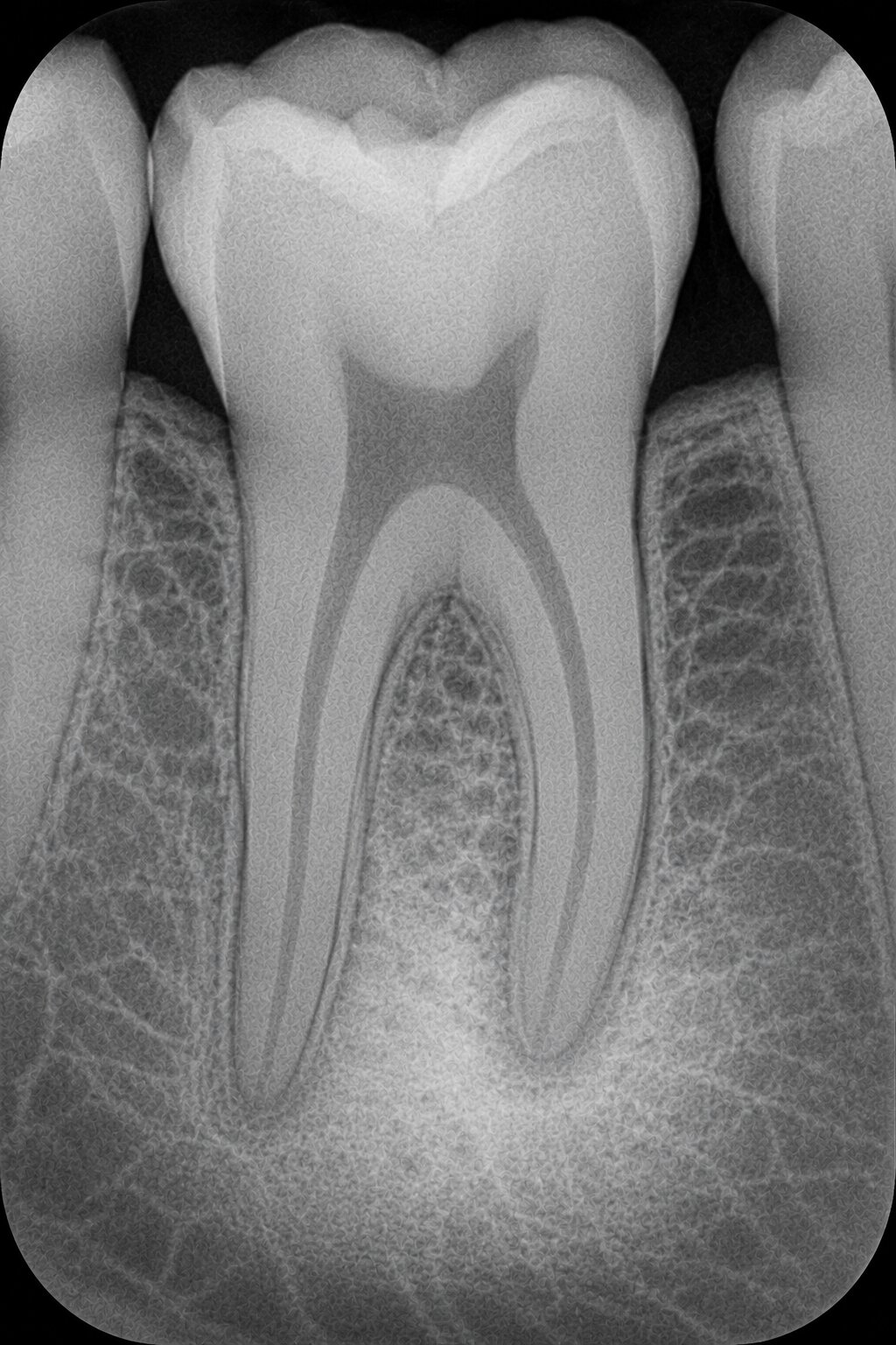
That denser, whiter patch near the root tip is sclerotic (condensing) bone — more mineral, so it blocks more beam. No dark border: it's dense bone, not a cavity or cyst.
The dark end, for real
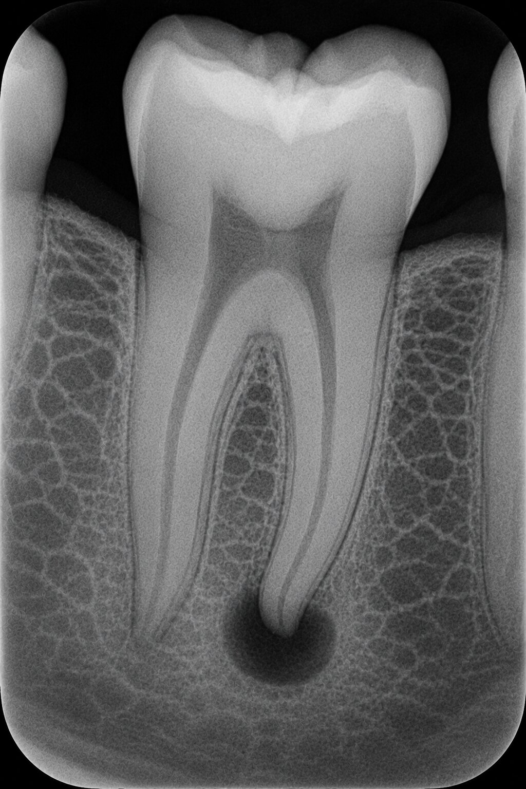
Periapical lesion
That dark balloon at the root tip is a periapical radiolucency — bone destroyed by infection. Rounded, dark, and centered on the apex — the opposite of the dense patch above.
5The traps
The look-alikes that fool people.
Three findings sit right next to "caries" or "error" on the exam and catch people who only memorized definitions. Learn the mimic, not just the term:
"radiolucency mistaken for caries"→Cervical burnout — a normal dark band at the neck of the tooth
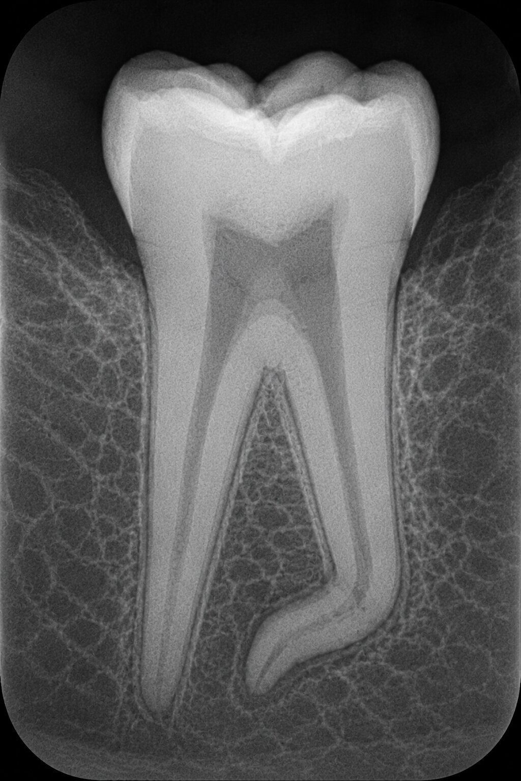
"abnormally bent or curved root"→Dilaceration — a real anomaly, not an angulation error
"bilateral finding, both sides"→Normal anatomy — symmetry usually = a landmark, not pathology
"dense white artifact / very bright"→Metal — restoration or metallic partial denture
The caries impostor
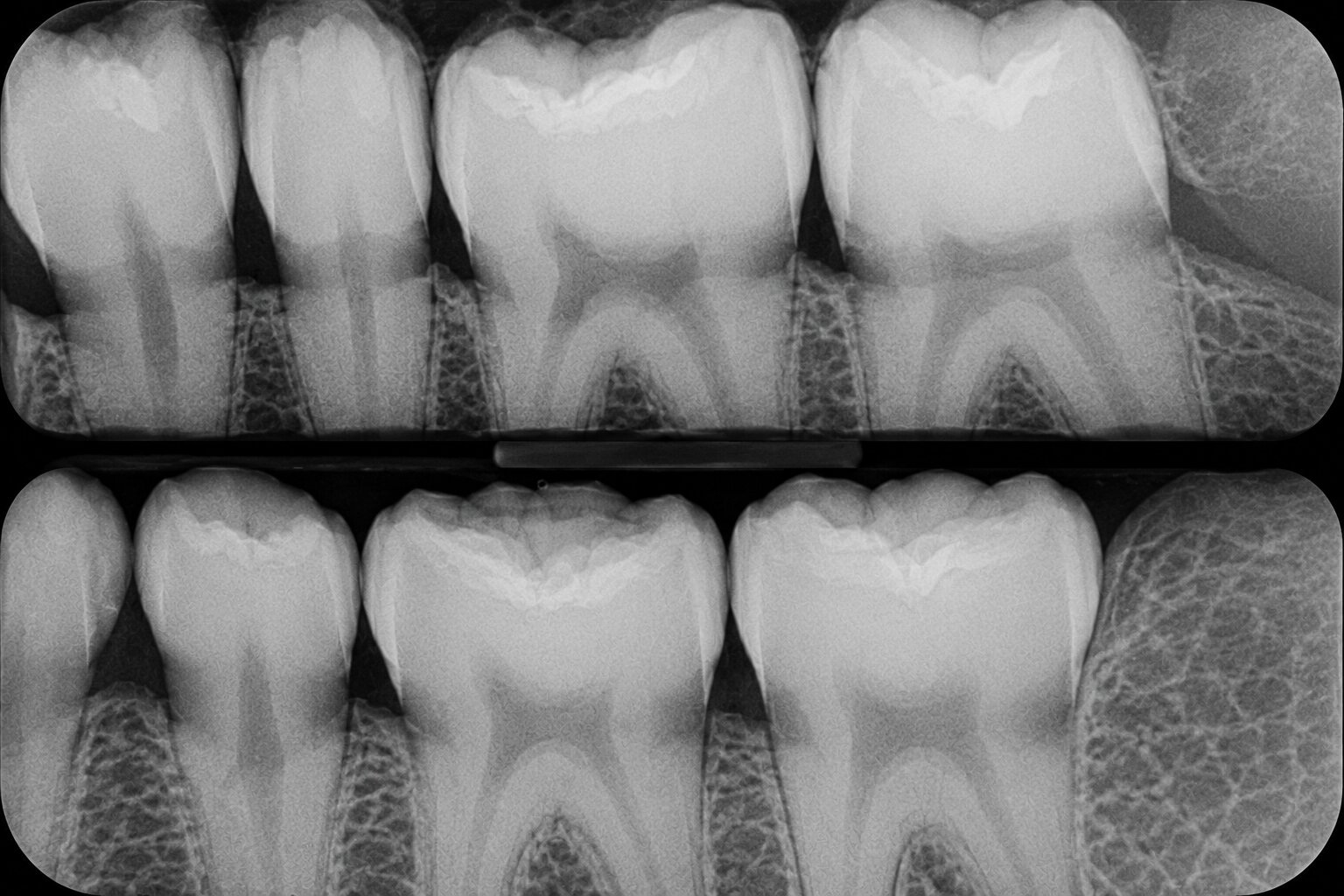
Cervical burnout
That soft dark collar sits at the tooth's neck, below the enamel, above the bone — and repeats tooth after tooth. Normal: the neck is just thinner there. A single dark cavity would be caries; this repeating band is burnout.
Dilaceration, in the flesh
Dilaceration
The root runs straight, then bends sharply near the tip. An anatomical anomaly — the root really is curved — not an angulation error, so repositioning won't "fix" it.
6Check yourself
Which of the following lesions appears radiopaque?
Sclerotic bone. Condensing/sclerotic bone is denser than normal bone, so it reads light (radiopaque). Cysts, abscesses, and most other lesions have lost mineral and read dark (radiolucent).
6Check yourself
Which radiolucency may be mistaken for caries?
Cervical burnout. It's a normal dark band at the cervical "neck" of the tooth (between enamel and bone) that can look like root caries. Calculus, sclerotic bone, and metal are all radiopaque, so they can't be mistaken for a dark cavity.
6Check yourself
How do most carious lesions appear on an image?
Radiolucent. Decay removes mineralized tooth structure, so less beam is blocked and the area reads dark. That's why a "dark spot" between teeth on a bitewing raises the flag for interproximal caries.
Recap
The whole thing in 5 lines.
✓Radiopaque = light = dense (blocked the beam). Radiolucent = dark = less there (let the beam through).
✓Brightest to darkest: metal → enamel → bone/dentin → caries → pulp/PDL → cysts/abscess → air.
✓Most carious lesions read radiolucent — decay removes mineral, so more beam gets through.
✓Cervical burnout mimics caries; dilaceration mimics an angulation error. Neither is what it looks like.
✓A finding on both sides is usually normal anatomy, not pathology.
Two words carry most of the image questions. Opaque is light, lucent is dark — say it until you don't have to think.
Then the exam's favorite move — a dark band that isn't caries, a bent root that isn't an error, a bright spot that's just metal — stops working on you. You're not memorizing pictures; you're reading density.