Before you can take a safe image, you need to know the equipment in your hand. The good news: there are only a handful of parts that matter for the exam — and each one has a clear job.
7 parts that matter~9 min★ The exam tests parts by their JOB, not just their name
▶ Watch · 2:08
Watch first — every part of the x-ray machine, including the PID, in under two and a half minutes.
1What & why
Think of it as a camera for teeth.
The dental x-ray unit has three parts you handle (tubehead, PID, control panel), two parts inside that actually make the x-rays (cathode and anode), and two parts that protect the patient (aluminum filter and collimator).
You already understand a camera — point it, press the button, it captures an image. The tubehead is the camera body. The PID is the lens you point at the subject. The control panel is the settings dial. Deep inside the tubehead, the cathode and anode are the "flash" — where the energy is actually created. You don't need to memorize a parts catalog — you need to know what each part does, because that's exactly how the exam asks.
2The parts you handle
The three outside parts.
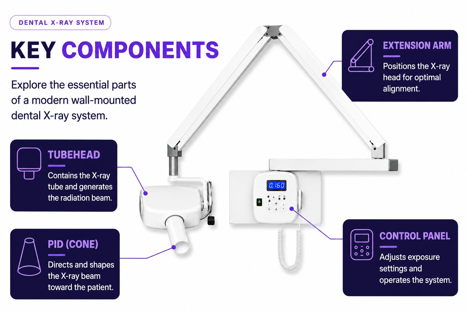
These are the parts you physically touch in the operatory. Here's each one and its job:
The partIts job
📷TubeheadThe sealed metal head of the machine. Inside it sits the x-ray tube, surrounded by insulating oil.
→
Houses the x-ray tubethe metal head that holds everything that makes the beam
🎯PIDPosition Indicating Device, also called the "cone." The open-ended tube you aim at the patient. (cone)
→
Directs & aims the beampoints the x-rays at the receptor; if it's off, you get a cone-cut — and a longer PID restricts the beam more than a shorter one
🎛️Control panelThe buttons/screen where you set exposure factors and press to expose.
→
Sets kVp, mA & timewhere you choose the exposure settings and fire
(There's also an extension arm — the jointed arm that positions the tubehead. It's rarely tested by name; just know it holds and moves the tubehead.)
On a real machine: the tubehead (with the PID/cone pointing down), the extension arm holding it to the wall, and the control panel. These are the parts you handle every appointment.
3How x-rays are made
Inside the tubehead.
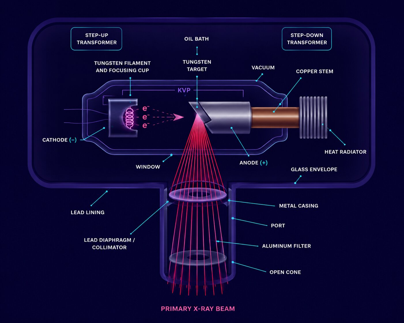
Open the tubehead and you'd find the x-ray tube — a sealed glass vacuum tube with two ends. This is where the actual x-rays are born:
Inside the tube: the cathode (−) releases electrons that race across the vacuum and strike the anode's tungsten target (+), producing x-rays. The beam then passes through the aluminum filter and collimator before exiting the cone toward the patient.
So the order is always: the cathode (negative) heats up and boils off electrons → those electrons race across the vacuum → they slam into the anode's tungsten target (positive) → that collision releases x-rays, which exit toward the patient. The vacuum matters: it lets the electrons fly across without bumping into air.
The partIts job
−CathodeThe negative (−) side. Its tungsten filament heats up and releases a cloud of electrons.
→
Source of electronsheated filament boils off electrons
+AnodeThe positive (+) side. Holds the tungsten target the electrons crash into.
→
Target — x-rays made hereelectrons strike the tungsten target → x-rays
4Make it stick
Cathode = down / negative
Cathode sounds like "the descent" — the negative start where electrons come from. The anode is the + target they go to. Electrons always travel cathode (−) → anode (+).
One exam term worth knowing: when the cathode filament heats up and releases its cloud of electrons, that's called thermionic emission — "thermionic" simply means heat releasing charged particles.
5The wave itself
X-rays are light you can't see.
X-rays are electromagnetic radiation — the same family as visible light and radio waves, just far higher energy. Three properties describe any wave in that family:
PropertyWhat it means for x-rays
〰️WavelengthThe distance between one wave peak and the next.
→
Very shortshorter than visible light — that short wavelength is what lets x-rays penetrate tissue
📶FrequencyHow many wave cycles pass a point per second.
→
Very highshort wavelength always means high frequency — the two move in opposite directions
⚡VelocityThe speed the wave travels.
→
Speed of lightthe same for every wavelength — x-rays, visible light, and radio waves all travel at roughly 3×10⁸ m/s in a vacuum
The one relationship worth remembering: wavelength and frequency move in opposite directions — shorter wavelength = higher frequency = higher energy = more penetrating. That's exactly why x-rays (short wavelength) pass through soft tissue while visible light (much longer wavelength) doesn't.
6Protecting the patient
The two parts the exam loves.
Both of these tie straight into radiation protection — each one makes the beam safer for the patient:
The partHow it protects the patient
🛡️Aluminum filterA thin aluminum disc in the path of the beam, inside the tubehead.
→
Removes weak photonsfilters low-energy x-rays that would only add dose, not image — machines at 70 kVp+ need 2.5 mm of aluminum
⏹️CollimatorA lead plate with an opening; a rectangular collimator shapes the beam to the receptor size.
→
Restricts beam sizelimits the beam to the area being imaged — no wider than 2.75 in. at the patient's skin
Easy way to keep them straight: the filter changes the beam's quality (takes out the weak rays); the collimator changes the beam's size (makes it smaller). Both lower patient dose.
The PID length matters here too: a longer PID restricts the beam more than a shorter PID. The beam spreads (diverges) as it travels, so the extra length gives it less room to fan out by the time it reaches the patient — a tighter beam, less scattered tissue exposure, and lower dose.
7Beat the exam
How DANB will actually ask this.
Notice the pattern — the exam describes a job and asks for the part:
"where x-rays are produced"→Anode (tungsten target)
"source of electrons"→Cathode (filament)
"reduces patient dose by removing weak rays"→Aluminum filter
"restricts the size of the beam"→Collimator
"directs the beam toward the receptor"→PID
"which PID length restricts the beam more"→longer PID — less beam divergence
"compare x-rays to visible light"→x-rays: shorter wavelength, higher frequency, higher energy
8Check yourself
In the dental x-ray tube, x-rays are actually produced when high-speed electrons strike the:
Tungsten target of the anode. Electrons come FROM the cathode (−) and strike the anode's tungsten target (+), where the collision produces x-rays.
8Check yourself
The aluminum filter in the tubehead reduces patient dose primarily by:
Removing low-energy photons. Those weak rays can't reach the receptor — they'd only deposit dose in the patient. Restricting beam SIZE is the collimator's job, not the filter's.
8Check yourself
Compared to visible light, x-rays have a:
Shorter wavelength and higher frequency. The two always move in opposite directions — the shorter the wavelength, the higher the frequency and the more energy the wave carries, which is why x-rays penetrate tissue and visible light doesn't.
Recap
The whole thing in 6 lines.
✓Tubehead holds the tube · PID aims the beam (longer PID = more beam restriction) · control panel sets kVp/mA/time.
✓Cathode (−) makes electrons → they strike the anode (+) tungsten target → x-rays.
✓Cathode = the negative descent (where electrons come from).
✓X-rays are EM radiation with a short wavelength and high frequency, traveling at the speed of light — short wavelength = high energy = more penetration.
✓Aluminum filter removes weak rays (beam quality) — lowers dose. At 70+ kVp, need 2.5 mm.
✓Collimator restricts beam size (no wider than 2.75 in. at the skin) — lowers dose.
That's the whole machine. On test day, you won't hunt for the part — you'll name it by its job.
When a question describes what a component does — makes electrons, produces x-rays, removes weak rays, restricts the beam — you'll match it to the right part on reflex. That's mastery, not memorization.